The eyes have long been called “windows to the soul” — and a growing body of research suggests they may also be a window into one of the earliest, most elusive signs of Alzheimer’s disease: amyloid buildup in the brain, years before symptoms appear.
The Discovery: Amyloid Plaques in the Retina
Amyloid plaques — clumps of misfolded protein that interfere with brain cell function — are one of the defining hallmarks of Alzheimer’s disease. For decades, detecting them required either an expensive PET brain scan or an invasive spinal tap. That may be starting to change.
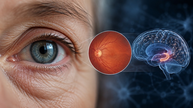
Researchers at the University of California San Diego School of Medicine found that the same amyloid plaques that accumulate in the brains of Alzheimer’s patients also build up in the retina — the light-sensitive tissue at the back of the eye. Because the retina is essentially an extension of the central nervous system, connected directly to the brain via the optic nerve, it offers a rare, non-invasive glimpse into processes normally hidden deep inside the skull.
What the Study Found
The UC San Diego team, led by Dr. Robert Rissman, professor of neurosciences and director of the Biomarker Core for the Alzheimer’s Disease Cooperative Study, compared retinal imaging with brain amyloid PET scans in participants from two major dementia research programs.
The results, published in the journal Alzheimer’s & Dementia in August 2021, showed a correlation: participants with visible amyloid spots on retinal imaging also tended to show higher levels of amyloid on their brain scans.
It’s worth being upfront about the scale of this particular study, because the original reporting undersold it: this was a small, cross-sectional screening analysis involving just eight patients.
Dr. Rissman himself was careful to frame it that way. “This was a small initial dataset,” he said, while adding that the findings were “encouraging because they suggest it may be possible to determine the onset, spread and morphology of Alzheimer’s disease — a preclinical diagnosis — using retinal imaging, rather than more difficult and costly brain scans.” He noted that the logical next step is a larger study to more thoroughly establish the relationship between retinal and cerebral amyloid over time.
This Wasn’t an Isolated Finding
What makes the UC San Diego results more credible is that they don’t stand alone. Earlier foundational research, led by Dr. Maya Koronyo-Hamaoui and colleagues, examined retinal tissue directly and found a striking 4.7-fold increase in amyloid-beta plaques in the retinas of confirmed Alzheimer’s patients compared with healthy controls, along with measurable neuron loss.
That same team demonstrated that retinal amyloid patterns closely mirrored the amyloid patterns found in the brain’s visual cortex.
Since then, other research groups have explored complementary approaches — including retinal blood vessel changes and nerve fiber layer thinning as possible early indicators of preclinical Alzheimer’s, and more recent work using hyperspectral retinal imaging to distinguish dementia from non-dementia patients across a larger sample of participants. Taken together, these studies point in a consistent direction, even though each individual study still has real limitations in sample size or design.
Why This Matters — And Why It’s Not There Yet
If retinal imaging for Alzheimer’s risk is eventually validated in large, well-controlled trials, the implications are significant. A basic eye exam is inexpensive, painless, quick, and already part of routine healthcare for many older adults — a dramatic contrast to PET scans, which can cost thousands of dollars and expose patients to radiation, or spinal taps, which are invasive and uncomfortable.
Earlier detection also matters clinically: catching amyloid buildup before cognitive symptoms emerge is exactly the window in which future treatments and lifestyle interventions are expected to be most effective.
That said, retinal amyloid imaging is not yet a validated diagnostic tool, and it isn’t something available at a standard optometrist visit today. The technology used in these studies — specialized imaging combined with amyloid-binding markers like curcumin — is still confined to research settings. No one should interpret a routine eye exam as an Alzheimer’s screening at this stage.
The Bottom Line
The idea that Alzheimer’s disease leaves detectable fingerprints in the eyes, years before memory symptoms appear, is one of the more promising directions in early-detection research. But “promising” is the operative word — the studies so far, including the widely cited eight-patient UC San Diego analysis, are early-stage and need to be confirmed in much larger populations before retinal imaging becomes a real clinical tool.
For now, if you’re concerned about Alzheimer’s risk — for yourself or a family member — the established path is still a conversation with a neurologist about cognitive screening and, where appropriate, brain imaging or biomarker testing.
References
- UC San Diego Health — “Eyes Provide Peek at Alzheimer’s Disease Risk”
- EurekAlert! (AAAS) — “Eyes provide peek at Alzheimer’s disease risk”
- Ngolab, J., et al. “Feasibility study for detection of retinal amyloid in clinical trials: The A4 study.” Alzheimer’s & Dementia: Diagnosis, Assessment & Disease Monitoring, 2021.
- Koronyo, Y., et al. “Retinal amyloid pathology and proof-of-concept imaging trial in Alzheimer’s disease.” JCI Insight, 2017.
- PMC — “Linking retinal amyloid imaging to cognition and hippocampal integrity”
- PMC — “Amyloid beta biomarker for dementia detection by hyperspectral ophthalmoscope images”